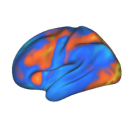
26) Some complex object categories, such as faces, have dedicated areas of cortex for processing them, but are also represented in a distributed fashion (Kanwisher – 1997, Haxby – 2001)
Early in her career Nancy Kanwisher used functional MRI (fMRI) to seek modules for perceptual and semantic processing. She was fortunate enough to discover what she termed the fusiform face area; an area of extrastriate cortex specialized for face perception.
This finding was immediately controversial. It was soon shown that other object categories also activate this area. Being the adept scientist that she is, Kanwisher showed that the area was nonetheless more active for faces than any other major object category.
Then came a slew of arguments purporting that the face area was in fact an ‘expertise area’. This hypothesis states that any visual category with sufficient expertise should activate the fusiform face area.
This argument is based on findings in cognitive psychology showing that many aspects of face perception once thought to be unique are in fact due to expertise (Diamond et al., 1986). Thus, a car can show many of the same perceptual effects as faces for a car expert. The jury is still out on this issue, but it appears that there is in fact a small area in the right fusiform gyrus dedicated to face perception (see Kanwisher’s evidence).
James Haxby entered the fray in 2001, showing that even after taking out the face area from his fMRI data he could predict the presence of faces based on distributed and overlapping activity patterns across visual cortex. Thus it was shown that face perception, like visual perception of other kinds of objects, is distributed across visual cortex.
Once again, Kanwisher stepped up to the plate. The following year she extended Haxby’s findings by showing that object categories can be recognized in distributed representations even with changes in viewpoint or exemplar. Importantly, she also showed that the face area carried special information about faces; it could be used to distinguish between faces and other categories (e.g., faces and bottles), but not two non-face categories (e.g., houses and bottles).
A series of other areas for specialized visual processing have also been found. In addition to the fusiform face area, Kanwisher also found the parahippocampal place area (for specialized processing of scenes) and the extrastriate body area (which is specialized for body part processing). Also, others have discovered the visual word form area (specialized for processing written words).
The visual word form area adds a special constraint to the other visual processing areas. While the other visual categories (faces, places, body parts) were all present in the ancestral environment and may have contributed to evolutionary pressures, word forms were not and could not.
Thus, the visual word form area could not have evolved but must rather be due to coincident conjunction of visual features that happens to be consistent across individuals. In other words, it must be something akin to an evolutionary spandrel (i.e., adaptive side-effect).
Applying this constraint on the other specialized processing areas suggests that they may have only peripheral roles in visual processing, as they are (as Haxby showed) only a small part of the larger visual processing stream.
Ultimately, the Kanwisher and Haxby debate illustrates another way (see previous post) in which the old distributed vs. localized function debate can be resolved. In this case it appears that the visual system is composed of a series of specialized regions dedicated to functions at differing levels of complexity working together to form distributed representations.
Note that these kinds of specialized regions are not restricted to visual processing. There is also a human vocalization area in auditory cortex (specialized for processing verbal and non-verbal human vocalizations; Belin et al., 2000). This suggests that insights gained in studying the visual processing system may generalize to all sensory modalities.
Implication: The mind, largely governed by reward-seeking behavior on a continuum between controlled and automatic processing, is implemented in an electro-chemical organ with distributed and modular function consisting of excitatory and inhibitory neurons communicating via ion-induced action potentials over convergent and divergent synaptic connections altered by timing-dependent correlated activity often driven by expectation errors. The cortex, a part of that organ organized via local competition and composed of functional column units whose spatial dedication determines representational resolution, is composed of many specialized regions forming specialized and/or overlapping distributed networks involved in perception (e.g., touch: parietal, vision: occipital), action (e.g., frontal), and memory (e.g., short-term: prefrontal, long-term: temporal), which depend on inter-regional connectivity for functional integration, population vector summation for representational specificity, dopamine signals for reinforcement learning, and recurrent connectivity for sequential learning.
[This post is part of a series chronicling history’s top brain computation insights (see the first of the series for a detailed description). See the history category archive to see all of the entries thus far.]
-MWCole
Kanwisher, N. (1997). The fusiform face area: a module in human extrastriate cortex specialized for face perception. Journal of Neuroscience, 17(11), 4302-4311.
Kanwisher, N., Yovel, G. (2006). The fusiform face area: a cortical region specialized for the perception of faces. Philosophical Transactions of the Royal Society B: Biological Sciences, 361(1476), 2109-2128. DOI: 10.1098/rstb.2006.1934
Haxby, J.V. (2001). Distributed and Overlapping Representations of Faces and Objects in Ventral Temporal Cortex. Science, 293(5539), 2425-2430. DOI: 10.1126/science.1063736

Great review of the FFA debate. Thanks for linking to your sources – I now have quite a few articles to read up on.